Noah Medical’s Jian Zhang on Galaxy II and the Future of Real-Time Robotic Bronchoscopy
04 September 2026 | Interaction | By Editor Robotics Business NEWS <editor@rbnpress.com>
In conversation with Robotics Business News, Jian Zhang discusses how Galaxy II advances imaging flexibility, robotic precision and real-time confirmation for earlier lung cancer diagnosis.
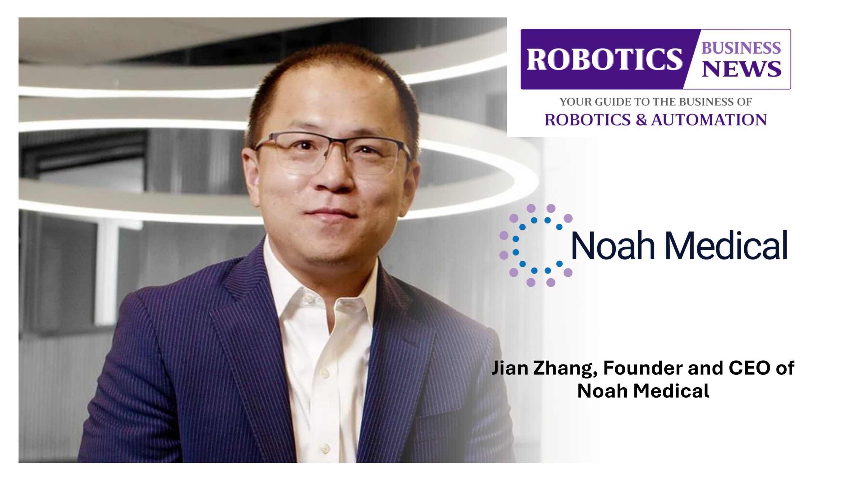
In conversation with Robotics Business News, Jian Zhang, Founder and CEO of Noah Medical, discusses the Galaxy II software update, its dual-mode imaging capabilities, enhanced robotic control and the company’s vision for expanding access to more accurate, efficient and earlier lung cancer diagnosis.
What were the key clinical and physician needs that drove the development of Galaxy II software, and how does it advance the Galaxy System?
The Galaxy II update came directly out of what physicians told us across more than 15,000 procedures worldwide. The original Galaxy System was built to solve a specific problem in robotic bronchoscopy. Even after two generations of navigation technology, roughly 20 percent of biopsies were still coming back inconclusive, and diagnostic yield had plateaued around 80 percent.1-2 The reason wasn’t that physicians couldn’t get close to a lesion. It was that they couldn’t confirm their tool was actually in it before taking the sample.
The Galaxy System closed that gap by integrating navigation with real-time imaging and confirmation of tool-in-lesion. The clinical results validated the approach, and adoption followed.
What we heard next was different. Physicians weren't asking us to fix something broken, they were asking for more optionality in a workflow that already worked. Three requests came up consistently: in a small subset of genuinely complex cases, they wanted higher-definition imaging without leaving the platform; they wanted more precise and predictable robotic control through difficult anatomy; and they wanted fewer manual steps during image acquisition, both for efficiency and for radiation safety.
The Galaxy II update answers all three concerns. Dual-Mode Imaging lets physicians move between the High-Definition DT mode (HD-DTTM Imaging) and Cone-Beam CT (CBCT) at any point in a procedure. An enhanced driving experience delivers increased speed, responsiveness, and maneuverability. Auto Capture automates image recording in sync with the C-arm sweep. The Galaxy II update adds the power of choice, control, and efficiency on a platform already delivering strong outcomes.
How does Dual-Mode Imaging, combining HD-DT imaging and CBCT, change the way physicians approach complex lung lesion biopsies?
It changes the conversation from a binary choice made before the case to a clinical judgment made during it.
Historically, imaging strategy was decided in advance by the equipment in the room. If a site is built around Cone-Beam CT, every case gets CBCT, including routine ones that don't need it, with the associated time and radiation cost. If a site doesn't have CBCT, the option isn't there for the complex cases where it would genuinely help.
Dual-Mode Imaging removes that constraint. HD-DT imaging uses a partial arc sweep and delivers the clarity most cases need quickly and at a fraction of the radiation dose. CBCT imaging uses a wider rotation, a minimum of 140 degrees, and provides the best available image quality for the most challenging anatomy. Both run on the same platform, and physicians can move between them at any point in the procedure without a reset and without losing focus.
What makes that possible is that the imaging data is embedded in the robot rather than borrowed from a separate imaging system. Because we own the raw image, we can keep refining and optimizing reconstruction specifically for lung biopsy, and that is what HD-DT imaging is. Not all digital tomosynthesis is created equal: most is general-purpose imaging applied to the lung, ours is built for this procedure and this target. Owning the image also gives physicians control beyond the choice of mode. They set the sweep angles and shape acquisition around the lesion in front of them, optimizing for the clearest view at the lowest dose rather than accepting a fixed protocol.
Practically, a physician can navigate confidently with HD-DT imaging on the majority of cases and reach for CBCT imaging only when added definition genuinely changes the plan. The decision sits with the physician, case by case, rather than with the equipment in the room. And because the Galax System has the broadest C-arm compatibility in the category, most hospitals can use imaging equipment they already own.
CT-to-Body Divergence and Tool-Tissue Divergence remain major challenges in bronchoscopy. How does the Galaxy II update address these issues in real time?
These are two distinct problems requiring two distinct solutions, a distinction the field doesn’t always make. We organize our approach around it: scope targeting corrects CT-to-Body Divergence, tool targeting corrects Tool-Tissue Divergence.
CT-to-Body Divergence occurs because the preoperative CT is a static snapshot of a lung that is no longer in the same configuration once the patient is under anesthesia and being ventilated. Differences in lung volume, atelectasis, respiratory and cardiac motion, and simple time elapsed since the scan all contribute. The Galaxy System addresses this with intraprocedural 3D imaging, HD-DT imaging or CBCT imaging, that updates lesion localization to the patient’s anatomy at that moment, plus virtual targeting guidance for scope positioning.
Tool-Tissue Divergence is a different failure mode. Even with the catheter correctly positioned, the biopsy tool can deviate from its intended trajectory as it exits the working channel and advances through tissue. Catheter flexibility, airway-wall resistance, tissue compliance, and deflection of the instrument itself all play a role. A great deal of diagnostic yield is lost here, and better navigation alone cannot fix it. The Galaxy System addresses it with an always-on camera that lets physicians verify tool approach and avoid critical structures, and with augmented fluoroscopy that overlays the lesion location on live fluoroscopy so physicians can see center strike in real time and adjust as the tool moves.
The Galaxy II update strengthens both. Lesion Focus reduces disruptive imaging artifacts and delivers a cleaner view of the target lesion and clearer confirmation of tool-in-lesion. The enhanced precision controls give physicians a finer command during exactly the maneuvers where divergence is most likely. Physicians can see the lesion, see the tool, and confirm the relationship between them before committing to a biopsy.
The Galaxy II update is designed to give physicians greater control over imaging decisions. How does this flexibility translate into improved procedural efficiency and confidence?
Efficiency and confidence come from the same source: matching imaging to the case rather than applying the same approach to every case.
On efficiency, the numbers are meaningful. Published data across roughly 700 procedures shows about 22 minutes shorter in-room time per case compared to CBCT-dependent workflows.3 That improvement comes from not leaving the room for spins, no cloud-based biopsies, no separate C-arm imaging workflow, and no added confirmation spins. Over a full day that can mean one more procedure, and we see roughly 25 percent higher utilization than competing platforms.
On confidence, it's about not having to compromise, and about being free to image as often as the case actually calls for. In the MATCH 2 study published in 2026, concordance between Galaxy’s HD-DT imaging and CBCT was 96.7 percent, so physicians know HD-DT imaging is delivering the information they need in the overwhelming majority of cases, and CBCT is available at any point if the anatomy proves more complex than expected. Just as important, because physicians control which modality they use and when, and because HD-DT imaging delivers up to 10 times lower dose than a CBCT spin, taking another image costs far less than it does in a CBCT-dependent workflow. The physician confirm, adjust, and confirm again without weighing every spin against a dose budget, instead of talking themselves out of an image they would rather have.
Radiation exposure is an important consideration in image-guided procedures. How does the ability to use HD-DT for most cases help reduce radiation exposure for patients and clinical teams?
This is one of the clearest advantages of the dual-mode approach, and it comes down to physics.
HD-DT imaging uses a partial arc sweep, while CBCT imaging requires a minimum 140-degree rotation and is generally around 200-degrees with a high image rate. Fewer X-ray projections mean substantially less dose. In our internal comparison, a 100-degree HD-DT sweep delivered 6.2 mGy versus 78.4 mGy for a 200-degree CBCT spin on the same target. Across cases, that translates to up to 10 times lower radiation dose on average with HD-DT imaging compared to CBCT imaging.*
The significance is cumulative. A patient undergoing a workup for lung cancer goes through multiple imaging studies across their diagnostic pathway, and a physician performs these procedures every week for years. Reducing dose on the majority of cases meaningfully lowers cumulative exposure for both.
Auto Capture adds a second layer of protection. By synchronizing image recording with the C-arm sweep, it eliminates the manual steps that kept staff in the room during acquisition. Clinical staff can step out while the image is captured, reducing scatter exposure without slowing the case.
All of this makes the Galaxy System the only RAB (Robotic-Assisted Bronchoscopy) platform that truly aligns with the ALARA principles, using the lowest effective radiation dose for what a given case actually requires. Physicians reserve fuller imaging for situations where it genuinely changes the plan, rather than defaulting to it everywhere.
What improvements in navigation, reach, flexibility, and stability can physicians expect from Galaxy II update when treating patients with challenging anatomy?
The enhanced drive experience touches every single case. The Galaxy II update delivers increased speed, responsiveness, and maneuverability compared to the prior generation. That predictability matters most when working in the periphery, where small deviations compound quickly. Physicians can reach all 18 segments of the lung with confidence, maintain stable positioning while advancing tools, and make finer adjustments during the maneuvers that determine whether a biopsy lands on target.
Stability is also something physicians can see rather than infer. Augmented fluoroscopy places an overlay of the lesion location onto live fluoroscopy, so physicians can watch the scope hold its position in real time and see exactly how that position relates to the target, whether the scope is already in the lesion or still driving toward it. In challenging anatomy, where respiratory motion and airway-wall resistance can shift a catheter that looked settled a moment earlier, that continuous view confirms the scope is stable and on line before a tool is advanced.
The single-use design means every case also starts with a scope performing at its best. A reusable scope carries its history with it: 34 reprocessing steps between cases, cumulative wear on the articulation, and gradual degradation of optics and torque response that can quietly cost performance in the periphery.4 A Galaxy scope has no history. Physicians get the same responsiveness, articulation, and image quality on the last case of the day as on the first, and because there is no next case to protect it for, they can drive it and deliver therapy through it without concern about damage. Every Galaxy case starts with a new scope.
The Galaxy System has demonstrated strong diagnostic performance in studies such as MATCH 2 and at Cleveland Clinic. How do these results influence Noah Medical’s approach to further clinical adoption?
MATCH 2 Study was a prospective, multicenter trial with 31 patients and three operators, designed to show what experienced users can achieve. It produced a 96.7 percent diagnostic yield using ATS strict definitions, currently one of the most conservative standards in the field, along with 96.7 percent concordance between HD-DT imaging and CBCT imaging, a 3 percent pneumothorax rate, and a 46-minute median case time that included an additional CBCT spin required by the study protocol.
Read together, those two 96.7 percent numbers are what give physicians confidence. The yield tells them that when they can see the lesion, see the tool, and confirm the relationship between the two before sampling, the biopsy lands. That is the clearest evidence yet for real-time visualization: the always-on camera that lets a physician verify tool approach, and augmented fluoroscopy that shows center strike as the tool moves. The concordance figure tells them that level of confirmation does not depend on CBCT, because HD-DT imaging delivered the same read in essentially every case. And the 46-minute median case time, which included a protocol-mandated CBCT spin, tells them none of it costs time.
The Cleveland Clinic experience extends that picture from a controlled trial into everyday practice. Across 100 consecutive cases and 116 nodules averaging just 12.9 mm, physicians achieved 98 percent tool-in-lesion and a 90 percent strict diagnostic yield.5 MATCH 2 showed what real-time confirmation delivers in a prospective multicenter setting; the Cleveland Clinic series shows the same targeting and diagnostic performance holding up in a demanding, unselected population that included very small lesions. Consistency across both settings is what convinces physicians the results are reproducible rather than study-specific.
That is also what shapes where we focus. Physician confidence is the gating factor for adoption, and it matters most outside the academic centers, in the community programs where most patients actually live and where operators may be earlier in their robotic experience. Real-time confirmation closes that gap: the physician isn't inferring, they are looking at the tool in the lesion. One new robotics program went from 20 TTNA cases to 150 Galaxy cases with a single operator and no CBCT, more than 500 percent growth, 95 percent diagnostic yield in year one, and over $2 million in net new biopsy revenue. A platform conversion site went from 150 cases on a competing robot to 480 on the Galaxy System, with 23-minute average case times enabling three more procedures per day and a 12-point improvement in diagnostic yield.* Those stories are why we've concentrated on community hospitals building lung nodule programs alongside academic centers pushing the boundaries on complex nodules from screening.
Looking ahead, how do you see the Galaxy II update and Robotic-Assisted Bronchoscopy evolving to support earlier and more accurate lung cancer diagnosis?
The biggest opportunity ahead isn't a more sophisticated procedure in a handful of academic centers. It's access. Lung cancer screening is expanding and more nodules are being found earlier, but most of those patients are in community hospitals, and most of those hospitals have never had robotic bronchoscopy. Diagnosing patients earlier at any real scale depends on two things: getting the technology to where the patients actually are, and getting more operators comfortable performing the procedure.
We are positioned to do that in a way earlier-generation platforms are not, because the Galaxy System doesn't require additional complex capital. There is no Cone-Beam CT suite to build, no reprocessing infrastructure to buy or staff. The Galaxy System has the broadest C-arm compatibility in the category, most hospitals can use imaging equipment they already own. That takes the capital conversation, which has kept robotic bronchoscopy out of most community programs, off the table.
Access alone isn't enough if the procedure feels difficult. Confidence comes from seeing what is happening, which is what augmented fluoroscopy and the always-on camera provide: the lesion overlaid on live fluoroscopy, the tool visibly approaching it, and confirmation before anyone takes a sample. For an operator who is still building experience, that information is what shortens the learning curve and makes strong results reproducible instead of expert-dependent.
Because the imaging data is embedded and processed on board, we own the full imaging chain, and that is the foundation for what comes next. We can keep developing tools that simplify the procedure: more visualization to support confident diagnosis, and continued improvement in image quality without CBCT. Every gain there lands where it matters most: a program that cannot afford a CBCT suite still gets best-in-class image quality.
Ultimately, the measure that matters is whether patients are being diagnosed at a stage where the disease is still curable. That means more programs, more confident operators, and better imaging in the places that have gone without. Everything we're building is aimed at that goal.